Renal Blood Vessels Labeled - Renal Blood Vessels Labeled Renal Artery Doppler Sonographic Tendencies The Hepatic System Is Important Because It Collects Blood From The Intestine And Passes It To The Liver The Centre For - They cleanse the blood of toxins and balance the constituents of the circulation to homeostatic set points through the processes of filtration, reabsorption, and secretion.
Renal Blood Vessels Labeled - Renal Blood Vessels Labeled Renal Artery Doppler Sonographic Tendencies The Hepatic System Is Important Because It Collects Blood From The Intestine And Passes It To The Liver The Centre For - They cleanse the blood of toxins and balance the constituents of the circulation to homeostatic set points through the processes of filtration, reabsorption, and secretion.. The renal arteries form directly from the descending aorta, whereas the renal veins return 'cleansed' blood directly to the inferior vena cava. By a single renal artery, but its not uncommon to find multiple. You will remember from gross anatomy that the renal artery enters the hilus of the kidney, and divides successively into lobar, interlobar (these are difficult to identify with certainty in histological sections, but they are the large arteries among the pyramids that are upstream of the. Emerging from the hilum is the renal pelvis, which is formed from the major and minor calyxes in the kidney. Berandarenal blood vessels labeled / renal circulation alila medical images :
The renal arteries form directly from the descending aorta, whereas the renal veins return 'cleansed' blood directly to the inferior vena cava. The nephrons also function to control blood pressure (via production of renin), red blood cell production (via the hormone erythropoetin), and calcium. Renal artery, one of the pair of large blood vessels that branch off from the abdominal aorta (the abdominal portion of the major artery leading from the heart) and enter into each kidney. The blood supply to the kidneys is carried by the renal arteries, paired. The left renal artery is much shorter and arises slightly more superior to the right main renal artery.
Pin On Med from i.pinimg.com Because the kidney filters blood, its network of blood vessels is an important component of its structure and function. The blood supply to the kidneys is carried by the renal arteries, paired. By a single renal artery, but its not uncommon to find multiple. Renal vascular anatomy • the renal pedicle classically consists of a single artery and a single vein that enter the kidney via the renal hilum. Both kidneys are superiorly related to an adrenal gland, and posteriorly to rib 12, the diaphragm, psoas major, quadratus lumborum and transversus abdominis. A medial indentation (the hilum) is where the renal blood vessels, nerves, lymphatic vessels, and ureter enter and exit the kidney. The left renal artery is much shorter and arises slightly more superior to the right main renal artery. You will remember from gross anatomy that the renal artery enters the hilus of the kidney, and divides successively into lobar, interlobar (these are difficult to identify with certainty in histological sections, but they are the large arteries among the pyramids that are upstream of the.
The arteries, veins, and nerves that supply the kidney enter and exit at the renal hilum.
Blood vessels are tubes that run through the transport system in which blood is transported. Just before reaching the kidney, each renal artery divides into five segmental arteries, which provide blood to the various regions of the kidney. From these arterioles branch the afferent arterioles.each afferent arteriole divides into a capillary network. Renal hilar nodes and then lumbar nodes. Both kidneys are superiorly related to an adrenal gland, and posteriorly to rib 12, the diaphragm, psoas major, quadratus lumborum and transversus abdominis. Utilizing the kidney and nephron models, locate the following vessels: Renal blood vessels anatomy the kidneys are highly vascular and thus are equipped with vast and intricate networks of circulation in order to effectively cleanse and modify vast amounts of blood.the hilum permits the entry of the arterial blood flow via the renal artery.the renal artery then branches off creating the interlobular arteries.these then pass between the renal pyramids via the. The renal hilum is the entry and exit site for structures servicing the kidneys: Blood circulation into and out of the kidneys is highlighted with colored arrows. Renal arteries carry unfiltered blood from the aorta to the kidneys blood vessels labeled. The nephrons also function to control blood pressure (via production of renin), red blood cell production (via the hormone erythropoetin), and calcium. These give off a series of branches which enter the cortex as interlobular arterioles. Internal anatomy of the kidney use flagged pins to identify the following parts of the internal kidney cortex renal column medullary pyramid minor calyx major calyx renal pelvis ureter renal artery renal vein checkpoint 3 do not move on until your instructor has signed off on your flags!
The nephrons also function to control blood pressure (via production of renin), red blood cell production (via the hormone erythropoetin), and calcium. Renal hilar nodes and then lumbar nodes. The renal arteries branch off of the abdominal aorta and supply the kidneys with blood. Renal vascular anatomy • the renal pedicle classically consists of a single artery and a single vein that enter the kidney via the renal hilum. Oxygenated blood comes to the kidneys from the right and left renal arteries off the abdominal aorta.
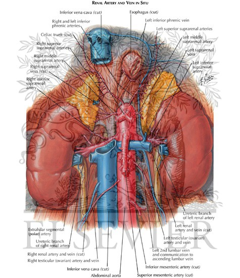
Renal Artery And Vein In Situ Renal Vasculature from www.netterimages.com Each kidney is typically fed. Search stock photos by tags Blood circulation into and out of the kidneys is highlighted with colored arrows. • the renal arteries arise from the aorta at the level of the intervertebral disk between the l1 and l2 vertebrae where the longer right renal artery passes posterior to the inferior vena cava (ivc). The renal arteries form directly from the descending aorta, whereas the renal veins return 'cleansed' blood directly to the inferior vena cava. The kidneys are important to the body's production of urine. Terms in this set (107) 1) the urinary system does all of the following, except that it a) excretes excess albumen molecules. The blood supply to the kidneys is carried by the renal arteries, paired.
Terms in this set (107) 1) the urinary system does all of the following, except that it a) excretes excess albumen molecules.
Blood vessels are an integral component of the circulatory system. You will remember from gross anatomy that the renal artery enters the hilus of the kidney, and divides successively into lobar, interlobar (these are difficult to identify with certainty in histological sections, but they are the large arteries among the pyramids that are upstream of the. Nephrons are the functional units of the kidney; They also play a role in regulating important components in the blood. Blood vessel names and roles are explained in this video, beginning with renal artery and ending with the cortical radiate arteries that serve the glomeruli. The renal cortex and medulla contain a complex network of blood vessels. • the renal arteries arise from the aorta at the level of the intervertebral disk between the l1 and l2 vertebrae where the longer right renal artery passes posterior to the inferior vena cava (ivc). The cushion of fat and the position of the kidney between the abdominal organs and muscles of the back protect it from trauma. Oxygenated blood comes to the kidneys from the right and left renal arteries off the abdominal aorta. Internal anatomy of the kidney use flagged pins to identify the following parts of the internal kidney cortex renal column medullary pyramid minor calyx major calyx renal pelvis ureter renal artery renal vein checkpoint 3 do not move on until your instructor has signed off on your flags! The nephrons also function to control blood pressure (via production of renin), red blood cell production (via the hormone erythropoetin), and calcium. Both kidneys are superiorly related to an adrenal gland, and posteriorly to rib 12, the diaphragm, psoas major, quadratus lumborum and transversus abdominis. The arteries, veins, and nerves that supply the kidney enter and exit at the renal hilum.
Renal hilar nodes and then lumbar nodes. The renal arteries form directly from the descending aorta, whereas the renal veins return 'cleansed' blood directly to the inferior vena cava. From these arterioles branch the afferent arterioles.each afferent arteriole divides into a capillary network. Renal blood vessels anatomy the kidneys are highly vascular and thus are equipped with vast and intricate networks of circulation in order to effectively cleanse and modify vast amounts of blood.the hilum permits the entry of the arterial blood flow via the renal artery.the renal artery then branches off creating the interlobular arteries.these then pass between the renal pyramids via the. Each kidney is typically fed.
Pin On My Schooling Journey from i.pinimg.com • the renal arteries arise from the aorta at the level of the intervertebral disk between the l1 and l2 vertebrae where the longer right renal artery passes posterior to the inferior vena cava (ivc). Because the kidney filters blood, its network of blood vessels is an important component of its structure and function. Renal artery (arteria renalis) the renal artery is a short paired artery that arises from the lateral aspect of the aorta. The arteries, veins, and nerves that supply the kidney enter and exit at the renal hilum. The interlobular arteries supply blood to the borders of the cortex and medulla, whereas the arcuate arteries diverge to form afferent arterioles that carry blood to the nephrons for filtration. Both kidneys are superiorly related to an adrenal gland, and posteriorly to rib 12, the diaphragm, psoas major, quadratus lumborum and transversus abdominis. Just before reaching the kidney, each renal artery divides into five segmental arteries, which provide blood to the various regions of the kidney. Blood vessels are an integral component of the circulatory system.
The renal cortex and medulla contain a complex network of blood vessels.
Renal blood flow is high relative to renal o2. They cleanse the blood of toxins and balance the constituents of the circulation to homeostatic set points through the processes of filtration, reabsorption, and secretion. Blood vessels (note outlines of red blood cells in. You will remember from gross anatomy that the renal artery enters the hilus of the kidney, and divides successively into lobar, interlobar (these are difficult to identify with certainty in histological sections, but they are the large arteries among the pyramids that are upstream of the. Supply blood to a functional segment of a kidney, divide into…. The interlobular arteries supply blood to the borders of the cortex and medulla, whereas the arcuate arteries diverge to form afferent arterioles that carry blood to the nephrons for filtration. The renal arteries branch off of the abdominal aorta and supply the kidneys with blood. Renal artery, segmental artery, interlobar artery, arcuate artery, cortical radiate artery, cortical radiate vein, arcuate vein, interlobar vein, and renal vein. Internal anatomy of the kidney use flagged pins to identify the following parts of the internal kidney cortex renal column medullary pyramid minor calyx major calyx renal pelvis ureter renal artery renal vein checkpoint 3 do not move on until your instructor has signed off on your flags! Berandarenal blood vessels labeled / renal circulation alila medical images : The nephrons also function to control blood pressure (via production of renin), red blood cell production (via the hormone erythropoetin), and calcium. Terms in this set (107) 1) the urinary system does all of the following, except that it a) excretes excess albumen molecules. Renal hilum renal pelvis renal sinus (with adipose) major calyx minor calyx renal.
The blood supply to the kidneys is carried by the renal arteries, paired blood vessels labeled. Renal blood flow is high relative to renal o2.


Posting Komentar
0 Komentar